Hi, Continue visiting your profile
user

Login

Email

Password

Create account
Forget password
Reset Password
you forgot your password?

Please enter your email address that you have used during registration:

AnyGenes

EPITHELIAL TO MESENCHYMAL TRANSITION: A KEY CELLULAR PROCESS

Epithelial to Mesenchymal Transition (EMT) is a dynamic and reversible biological process where epithelial cells lose their defining characteristics, such as cell-cell adhesion and apico-basal polarity, and acquire mesenchymal traits, including enhanced motility and invasiveness.

This transformation plays a pivotal role in diverse biological functions, including embryogenesis, wound healing, and pathological conditions like organ fibrosis and cancer progression.

AnyGenes® offers advanced qPCR arrays designed to study EMT-related gene expression and signaling pathways. Our high-quality tools empower researchers to investigate EMT’s molecular mechanisms, identify biomarkers, and explore its role in health and disease.

AnyGenes qPCR array for studying epithelial to mesenchymal transition (EMT) in cancer progression, fibrosis, and tissue regeneration.

Discover our advanced qPCR arrays for Epithelial to Mesenchymal Transition research.

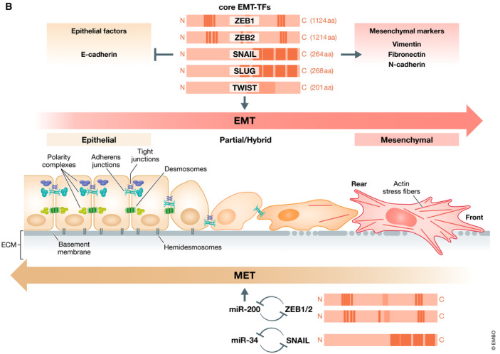
Epithelial-to-Mesenchymal-Transition-markers

(B) EMT is induced mainly by a set of transcription factors (EMT‐TFs) like ZEB1, ZEB2, SNAIL, SLUG and TWIST that differ in protein structure, size, and individual functions. All of them are repressors of epithelial factors like E‐cadherin and activate mesenchymal markers like Vimentin, Fibronectin or N‐cadherin. Epithelial cells displaying apical–basal polarity are held together by tight junctions, adherens junctions, and desmosomes and are anchored to the underlying basement membrane by hemidesmosomes. They express three different polarity complexes that together with the junctional molecules maintain epithelial cell polarity.

In the classical EMT, expression of EMT‐TFs leads to inhibition of major components of these epithelial structures and concomitantly activates the expression of genes associated with the mesenchymal state. Cells gain front–rear polarity, display actin stress fibers, become motile and acquire invasive capacities. Notably, tumor cells very rarely switch to a completely mesenchymal phenotype, but fluently convert between various intermediate states displaying certain mesenchymal features but keeping partial sets of epithelial characteristics. Further, EMT is a reversible process. Mesenchymal cells can revert to the epithelial state undergoing MET. An important role in the execution of MET is played by microRNAs of the miR‐200 and mir‐34 families that are regulated in double‐negative feedback loops with the EMT‐TFs ZEB1/2 and SNAIL, respectively, that serve to reinforce either the epithelial or the mesenchymal state.

MECHANISMS DRIVING EPITHELIAL TO MESENCHYMAL TRANSITION

EMT is orchestrated by key transcription factors that suppress epithelial markers (e.g., E-cadherin) and activate mesenchymal markers (e.g., vimentin, fibronectin):

  • Snail Transcription Factors: SNAIL1 and SNAIL2 repress epithelial genes while promoting mesenchymal ones.
  • Zinc-Finger Proteins: ZEB1 and ZEB2 regulate cellular plasticity during EMT.
  • TWIST Proteins: Drive the expression of genes linked to mesenchymal phenotypes.

SUBTYPES OF EPITHELIAL TO MESENCHYMAL TRANSITION

EMT is categorized into three distinct subtypes based on its biological context:

  • Type 1 EMT: Essential for embryonic development, tissue formation and organogenesis.
  • Type 2 EMT: Involved in wound healing, tissue regeneration, and fibrosis.
  • Type 3 EMT: Linked to cancer progression, enhancing metastasis and tumor invasiveness.

SIGNALING PATHWAYS DRIVING EMT

EMT is regulated by complex signaling pathways that integrate extracellular signals to control cellular behavior. Key pathways involved include:

  • TGF-β Signaling: A primary inducer of EMT, promoting the activation of transcription factors like SNAIL, ZEB, and TWIST.
  • Wnt/β-Catenin Pathway: Plays a crucial role in maintaining cellular plasticity during EMT, particularly in cancer and stem cell biology.
  • Notch Signaling: Influences EMT by modulating the expression of epithelial and mesenchymal markers.
  • PI3K/Akt Pathway: Regulates EMT through its impact on cell survival, migration, and invasion.

Understanding these pathways provides insight into the molecular events driving EMT and highlights potential therapeutic targets for diseases associated with aberrant EMT activity.

EMT IN CANCER PROGRESSION

In cancer biology, EMT plays a pivotal role in cancer progression by enabling epithelial cells to acquire invasive and migratory properties characteristic of mesenchymal cells. This process facilitates the spread of cancer cells from the primary tumor to distant sites, contributing to metastasis.

EMT also promotes chemoresistance by increasing cancer cell plasticity, enabling cells to survive therapeutic interventions and regenerate tumors. Understanding the mechanisms of EMT in cancer progression provides opportunities to develop targeted therapies aimed at mitigating metastasis and improving patient outcomes.

(1) Manfioletti G, Fedele M. Epithelial–Mesenchymal Transition (EMT) 2021. Int J Mol Sci. (2022);23(10):5848.
(2) Lachat C, et al. Epithelial to Mesenchymal Transition History: From Embryonic Development to Cancers. Biomolecules. (2021);11(6):782.
(3) Dudas J, et al. Epithelial to Mesenchymal Transition: A Mechanism that Fuels Cancer Radio/Chemoresistance. Cells. (2020);9(2):428.
(4) Marconi GD, et al. Epithelial-Mesenchymal Transition (EMT): The Type-2 EMT in Wound Healing, Tissue Regeneration and Organ Fibrosis. Cells. (2021);10(7):1587.
(5) Brabletz S, et al. Dynamic EMT: a multi‐tool for tumor progression. EMBO J. (2021);40(18):e108647.

EMT SIGNALING PATHWAY BIOMARKER LIST

Customize your own signaling pathways (SignArrays®) with the factors of your choice!
Simply download and complete our Personalized SignArrays® information file and send it at contact@anygenes.com to get started on your project.

You can check the biomarker list included in this pathway, see below:
Quitter la version mobile