hi continue visiting your profile
user

Login

Email

Password

Create account
Forget password
Reset Password
you forgot your password?

Please enter your email address that you have used during registration:

do you want to Log Out ?
AnyGenes

APOPTOSIS PATHWAY: KEY MECHANISMS IN CELL DEATH AND SURVIVAL

Apoptosis pathway, also known as programmed cell death, is a crucial process that maintains cellular homeostasis and eliminates damaged or unnecessary cells. Any disruption in apoptosis pathways can lead to various diseases, including cancer, autoimmune disorders, and neurodegenerative diseases. Understanding the Apoptosis Pathway provides insights into the mechanisms of cell death, which is essential for developing therapeutic approaches.

AnyGenes® Apoptosis qPCR Arrays: Powerful Tools for Advanced Research

Our Apoptosis Pathway qPCR arrays are designed to meet the needs of researchers exploring apoptosis in health and disease. With a comprehensive panel of apoptosis-related genes, these arrays allow for high-throughput analysis and precise gene quantification. AnyGenes products enable scientists to investigate apoptotic markers accurately, facilitating studies in oncology, immunology, and regenerative medicine.

Apoptosis Pathway qPCR Array by AnyGenes for analyzing gene expression in programmed cell death.
Apoptosis-Pathway

 
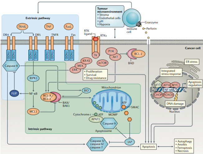
Overview of apoptosis signalling pathways and the effects of pro-survival signalling, immune cells and the tumour microenvironment
.

MAJOR PATHWAYS OF APOPTOSIS

The apoptosis pathway involves two main signaling mechanisms:

  • Extrinsic pathway: is initiated by the binding of extracellular ligands to specific death receptors on the cell surface. Key components include Death Receptors, members of the Tumor Necrosis Factor Receptor (TNFR) superfamily, including TNF receptor 1 (TNFR1), Fas (CD95), and TRAIL receptors, and Death-Inducing Signaling Complex (DISC).
  • Intrinsic pathway:The intrinsic pathway is primarily regulated by mitochondrial signals in response to internal stressors such as DNA damage, oxidative stress, heat, cytotoxic drugs or more specialized anti-cancer molecules. Key features include :

* Mitochondrial release: Stress signals lead to the permeabilization of the mitochondrial membrane, resulting in the release of pro-apoptotic factors like cytochrome c into the cytosol.

* Apoptosome Formation: Cytochrome c binds to Apaf-1 and procaspase-9, forming an apoptosome that activates caspase-9, which then activates downstream effector caspases.

  • Perforin/Granzyme Pathway: This pathway is utilized by cytotoxic T cells to induce apoptosis in target cells. It involves Perforin, forms pores in the target cell membrane, and Granzyme B. Entering through these pores, it activates caspases directly or initiates apoptosis via mitochondrial pathways.

MOLECULAR SIGNALING IN APOPTOSIS PATHWAY

Apoptosis involves a series of tightly regulated molecular signaling pathways that guide programmed cell death, which is essential for maintaining cellular balance and eliminating damaged cells. Beside intrinsic and extrinsic pathways, here are the key components and signaling pathways in apoptosis:

Crosstalk Between Pathways:

  • Key Link: Bid protein links extrinsic and intrinsic pathways.
  • Process: Caspase-8 from the extrinsic pathway cleaves Bid, promoting mitochondrial cytochrome c release to activate intrinsic signaling.

Execution Phase:

  •  Key Elements: Effector caspases (e.g., caspase-3) degrade cell components.
  • Process: Leads to DNA fragmentation, cell shrinkage, and formation of apoptotic bodies for clean cell removal.

Regulatory Proteins and Inhibitors:

  • Key Elements: IAPs (inhibit caspases), p53 (activates apoptosis in DNA-damaged cells), and survivin (prevents apoptosis in cancer cells).
  • Function: These regulators control apoptosis activation to maintain cellular balance and prevent unintended cell death.

DIFFERENCE BETWEEN NECROSIS AND APOPTOSIS PATHWAY

Necrosis and apoptosis are two distinct forms of cell death with different causes, processes, and consequences. Necrosis is an uncontrolled, often damaging form of cell death, while apoptosis is a controlled, programmed process that promotes cellular balance and health.

Cause and Triggers:

  • Necrosis: Often triggered by external injury, infection, toxins, or lack of blood supply, leading to accidental cell death.
  • Apoptosis: Triggered by internal signals as part of a regulated process, often to remove damaged, unneeded, or harmful cells.

Process and Mechanism:

  • Necrosis: Involves cell swelling, membrane rupture, and uncontrolled release of cell contents.
  • Apoptosis: Characterized by cell shrinkage, DNA fragmentation, and formation of apoptotic bodies, which are then safely engulfed by neighboring cells.

Outcome and Impact on Surrounding Tissue:

  • Necrosis: Causes inflammation in the surrounding tissue due to the uncontrolled release of cellular debris.
  • Apoptosis: Does not cause inflammation, as the cell contents are contained within apoptotic bodies and cleared by immune cells, maintaining tissue health.

Role in the Body:

  • Necrosis: Often harmful and typically occurs due to pathological conditions.
  • Apoptosis: Beneficial and part of normal development, immune response, and cellular turnover.

APOPTOSIS PATHWAY AND DISEASES

  • Apoptosis and cancer: cancer cells often evade apoptosis, allowing them to grow uncontrollably. Mutations in genes regulating apoptosis (e.g., p53 or Bcl-2) can prevent cell death and contribute to tumor formation.
  • Neurodegenerative diseases: excessive apoptosis in the brain can lead to the loss of neurons, contributing to diseases such as Alzheimer’s, Parkinson’s, and Huntington’s.
  • Autoimmune diseasesinsufficient apoptosis of immune cells may lead to the persistence of autoreactive cells, which can attack the body’s own tissues and lead to diseases like lupus and rheumatoid arthritis.

Other diseases:

  • Heart diseases: abnormal apoptosis of cardiac cells is implicated in heart diseases, particularly after a heart attack, where stress-induced apoptosis can worsen tissue damage. Excessive apoptosis in the heart can weaken cardiac function and contribute to heart failure.
  • Infectious diseases: some viruses, such as HIV, manipulate apoptotic pathways to evade the immune system or induce excessive apoptosis in immune cells, weakening the host’s defense.
  • Liver diseases: Chronic liver diseases, including hepatitis and cirrhosis, often involve apoptosis of hepatocytes (liver cells), contributing to progressive liver damage.
  • Developmental disorders: Apoptosis is essential in shaping organs and tissues during development. Disruption of apoptosis during embryonic development can result in malformations or developmental abnormalities.
(1) Jan R, Chaudhry GS. Understanding Apoptosis and Apoptotic Pathways Targeted Cancer Therapeutics. Adv Pharm Bull. (2019)1;9(2):205–218.
(2) Pfeffer CM, Singh ATK. Apoptosis: A Target for Anticancer Therapy. Int J Mol Sci. (2018)2;19(2):448.
(3) Adams JM, Cory S. The BCL-2 arbiters of apoptosis and their growing role as cancer targets. Cell Death Differ. (2018);25(1):27-36.
(4)  Carneiro BA, El-Deiry WS. Targeting apoptosis in cancer therapy. Nat Rev Clin Oncol. (2020);17(7):395-417.
(5) Kashyap D et al. Intrinsic and extrinsic pathways of apoptosis: Role in cancer development and prognosis. Adv Protein Chem Struct Biol. (2021);125:73-120.(6) Abdulhussein D et al.  Apoptosis in health and diseases of the eye and brain. Adv Protein Chem Struct Biol. (2021);126:279-306.
(7)
 Carneiro BA, El-Deiry WS. Targeting apoptosis in cancer therapy. Nat Rev Clin Oncol. (2020);17(7):395-417.
(8)
 Neophytou CM et al. Apoptosis Deregulation and the Development of Cancer Multi-Drug Resistance. Cancers (Basel). 2021 Aug 28;13(17):4363.

APOPTOSIS SIGNALING PATHWAY BIOMARKER LIST

Customize your own signaling pathways (SignArrays®) with the factors of your choice!
Simply download and complete our Personalized SignArrays® information file and send it at contact@anygenes.com to get started on your project.

You can check the biomarker list included in this pathway, see below:
Quitter la version mobile