Hi, Continue visiting your profile
user

Login

Email

Password

Create account
Forget password
AnyGenes

ENDOTHELIAL TO MESENCHYMAL TRANSITION (EndMT): A KEY CELLULAR PROCESS

Endothelial to Mesenchymal Transition (EndMT) is a complex biological process where endothelial cells detach from the endothelium and acquire mesenchymal cell characteristics. This transition involves significant changes in cell morphology, function, and molecular profile, enabling cells to migrate and contract.

Accelerate EndMT Research with AnyGenes®

AnyGenes® offers advanced qPCR arrays specifically designed to analyze the gene expression changes associated with EndMT. These tools provide unmatched sensitivity and precision, enabling researchers to study signaling pathways, cellular plasticity, and disease mechanisms. Whether investigating fibrosis, cancer, or cardiovascular conditions, AnyGenes® solutions empower scientists to unlock critical insights and drive innovation in genomic research.

Discover AnyGenes qPCR array for analyzing Endothelial to Mesenchymal Transition (EndMT) pathways, enabling precise insights into cell signaling and disease mechanisms.

Discover our advanced qPCR arrays for Endothelial to Mesenchymal Transition research.

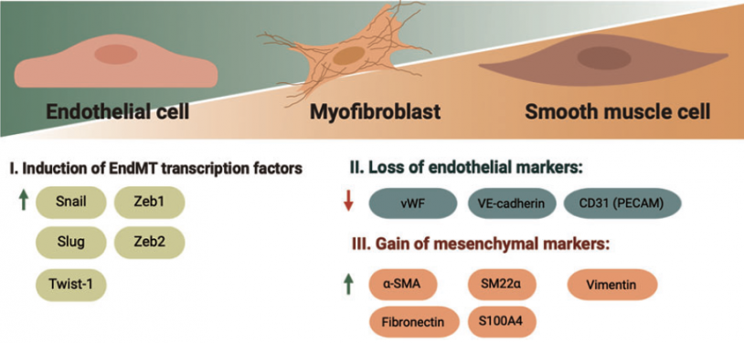
Endothelial-To-Mesenchymal Transition Progression

Sequence of events in the course of EndMT progression. Induction of EndMT-associated transcription factors Snai1, Snai2, Twist-1, Zeb1, and Zeb2 results in progressive loss of endothelial markers PECAM-1 and VE-cadherin, and gain of mesenchymal markers vimentin, fibronectin, SM22α, and α-SMA. α-SMA, alpha-smooth muscle actin; EndMT, endothelial-to-mesenchymal transition; PECAM-1, platelet endothelial cell adhesion molecule; SM22α, transgelin; Snai1, Snail; Snai2, Slug; VE-cadherin, vascular endothelial cadherin

KEY FEATURES OF ENDOTHELIAL TO MESENCHYMAL TRANSITION

  • Phenotypic Plasticity:
    EndMT enables endothelial cells to lose their endothelial-specific characteristics (e.g., polarity and barrier function) and acquire mesenchymal traits like motility and contractility.
  • Signaling Pathway Activation:
    • TGF-β Pathway: The primary driver of EndMT, particularly through SMAD-dependent signaling.
    • Other Pathways: Notable contributors include Wnt, Notch, and PI3K/Akt, which influence cellular transitions and promote mesenchymal phenotypes.
  • Biomarker Expression Shifts:
    • Downregulated Markers: VE-cadherin, CD31, and von Willebrand Factor (vWF).
    • Upregulated Markers: α-Smooth Muscle Actin (α-SMA), vimentin, fibronectin, and collagen types I and III.
  • Cytoskeletal Remodeling:
    • Significant reorganization of the cytoskeleton occurs to facilitate cell migration and contraction, crucial for tissue remodeling and repair.
  • Triggers and Stimuli:
    • Pathological Stimuli: Inflammation, oxidative stress, hyperglycemia, and low shear stress.
    • Physiological Contexts: Embryonic development, angiogenesis, and wound healing.
  • Dual Role in Health and Disease:
    • Physiological Role: Essential in vascular development and tissue repair.
    • Pathological Implications: Contributes to fibrosis, cancer progression, cardiovascular diseases, and chronic inflammation.

MECHANISMS OF ENDOTHELIAL TO MESENCHYMAL TRANSITION

EndMT is a process where endothelial cells transform into mesenchymal-like cells. This involves several key signaling pathways and factors:

  1. TGF-β Signaling Pathway: TGF-β activates intracellular pathways (SMAD and non-SMAD), driving mesenchymal markers like α-SMA and vimentin while suppressing endothelial markers.
  2. Wnt/β-Catenin Pathway: Wnt signaling activates β-catenin, enhancing mesenchymal gene expression and working synergistically with TGF-β to promote EndMT.
  3. Notch Signaling: Notch influences transcription factors like Snail and Twist, supporting the mesenchymal phenotype in collaboration with TGF-β.
  4. Hypoxia and Oxidative Stress: Low oxygen and reactive oxygen species (ROS) activate HIFs and MAPK/NF-kB pathways, driving EndMT, especially in fibrosis and cancer.
  5. Epigenetic Regulation: DNA and histone modifications alter gene expression, promoting mesenchymal markers and silencing endothelial ones.
  6. Mechanical Stress and ECM Interactions: Low shear stress and changes in the extracellular matrix (ECM) help initiate EndMT by altering cell behavior and reinforcing mesenchymal traits.
  7. Inflammatory Cytokines and Growth Factors: Cytokines like TNF-α and IL-1, as well as growth factors like VEGF, induce EndMT by promoting mesenchymal gene expression and disrupting endothelial cell junctions.

ENDMT IN PATHOPHYSIOLOGY

The mechanisms of EndMT are not only fundamental in developmental processes but are also implicated in several pathological conditions such as:

  • Fibrosis: EndMT contributes to the production of extracellular matrix proteins, leading to fibrosis in organs like the heart, lung, and kidneys.
  • Cancer: EndMT plays a role in the epithelial-mesenchymal transition (EMT) in cancer, contributing to tumor progression and metastasis.
  • Cardiovascular Diseases: In atherosclerosis and vascular remodeling, EndMT leads to the loss of endothelial function and the accumulation of mesenchymal cells in the vasculature.
  • Pulmonary Hypertension: EndMT in pulmonary endothelial cells contributes to the thickening of the vascular wall and vascular remodeling seen in pulmonary hypertension.

Studying EndMT is essential for understanding its dual roles in physiological and pathological processes. By leveraging AnyGenes® qPCR arrays, researchers gain access to a powerful toolset for decoding the complexity of this transition and its implications for health and disease.

Explore AnyGenes® products today and elevate your EndMT research to the next level.

ENDMT AND TISSUE REGENERATION

In addition to its role in disease progression, EndMT is also critical in normal physiological processes like tissue repair and regeneration. During wound healing, endothelial cells undergo EndMT to contribute to fibrosis and tissue remodeling, facilitating the repair of damaged blood vessels. This cellular plasticity allows for the formation of new fibroblasts that support tissue repair and maintain vascular integrity.

However, uncontrolled EndMT in pathological conditions can lead to excessive fibrosis, hindering normal tissue function. Understanding the balance between EndMT’s beneficial and detrimental roles in tissue regeneration is key to developing therapeutic strategies for conditions like chronic wounds and organ fibrosis.

(1) Bischoff J.  Endothelial-to-Mesenchymal Transition. Circ Res. (2019);124(8):1163-1165.
(2) Gorelova A,  Endothelial-to-Mesenchymal Transition in Pulmonary Arterial Hypertension. Antioxid Redox Signal. (2021);34(12):891-914.
(3) Piera-Velazquez S, Jimenez SA.  Endothelial to Mesenchymal Transition: Role in Physiology and in the Pathogenesis of Human Diseases. Physiol Rev. (2019);99(2):1281-1324.
(4) Xu Y, Kovacic JC.  Endothelial to Mesenchymal Transition in Health and Disease. Annu Rev Physiol. (2023);85:245-267.
 

ENDOTHELIAL TO MESENCHYMAL TRANSITION BIOMARKER LIST

Customize your own signaling pathways (SignArrays®) with the factors of your choice!
Simply download and complete our Personalized SignArrays® information file and send it at [email protected] to get started on your project.

You can check the biomarker list included in this pathway, see below: