
Logout
If you want to log out click in LogOut


| Signaling Pathways | Product ref | Informations |
|---|---|---|
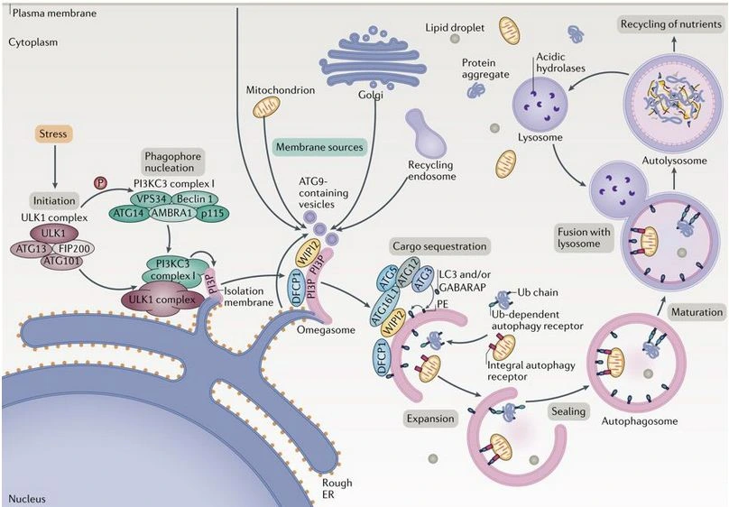
Autophagy | AT1H1 |

| Signaling Pathways | Product ref | Informations |
|---|---|---|
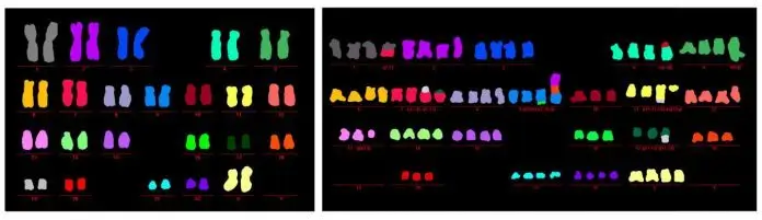
Autophagy | AT1M1 |
| Signaling Pathways | Product ref | Informations |
|---|---|---|
Autophagy | ******* |
Biomarker list in progress. |

| Signaling Pathways | Product ref | Informations |
|---|---|---|
Autophagy | ******* |
Biomarker list in progress. |