
Logout
If you want to log out click in LogOut


In transplantation, the antibody mediated rejection (ABMR) is currently a major thread for the long term graft survival. However the diagnosis of the ABMR remains still difficult and according to the last Banff classification the diagnosis of ABMR requires the evidence of current/recent antibody interaction with graft vascular endothelial cells: including one of following: linear c4d (a fragment of complement) staining in peritubular capillaries; or moderate microvascular inflammation; or increased endothelial injury related gene expression with thoroughly bio-statistically validated result. In the absence of easy access to transcriptomic analysis of graft samples, relying only on C4d staining or microvascular inflammation is clinically unsatisfactory.
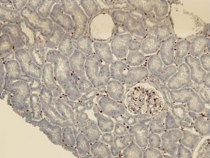
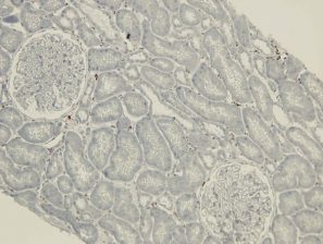
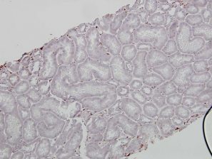
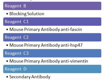
The ligation of HLA molecules on the vascular endothelial cells with corresponding antibodies and/or activation of complement following Antigen-Antibody complex formation can trigger cell stress response and by various signaling pathways it can affect the endothelial transcriptional profile and induce an up-regulation of molecules involved in inflammation, coagulation, cell motility and endothelial repair, a process reminiscing Endothelial to mesenchymal transition (EndMT). This EndMT process now can be detected on paraffin tissue by expression of fascin, hsp47 and vimentin (figures). The expression of these EndMT markers on the renal grafts is associated with the presence of DSA and predicts poor graft outcome.
fascin
hsp47
vimentin
No peri-tubular capillary EndMT marker staining in Normal renal grafts:
Peri-tubular capillary EndMT marker staining in renal grafts with antibody mediated rejection:
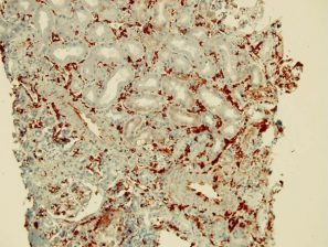
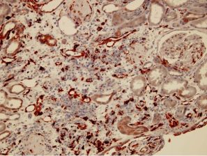
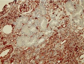
The EndMT Test, detects the endothelial cell injury and its implication in the antibody mediated rejection (see description above)
For more technical information, click here
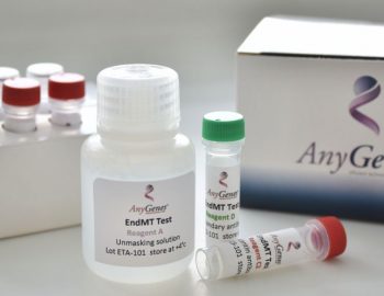
EndMT Test prices
| Reference type : EDMT-20 | Perfect Master Mix SYBRG® |
| Number of tests | Price before TAX |
|---|---|
| 20 | * |